A More Advanced Approach to Skin Cancer Screening
Early detection is critical in managing skin cancer. If you are considering a mole check or skin screening, the method used to examine and document your skin makes a meaningful difference.
At our dermatology clinic, we use an advanced AI-assisted mole mapping system combined with expert clinical examination to provide a more precise and structured way of monitoring your skin over time.
What happens during a mole check?
All mole checks begin with a full skin examination performed by a trained and experienced dermatologist with years of experience conducting skin cancer surveillance for high risk patients.
To ensure a thorough assessment, patients will be asked to undress to their underwear. This allows the doctor to examine the skin from head to toe, including less visible areas where skin cancers can still occur.
A dermatoscope (a specialised magnifying device) is used throughout the examination to assess the structure of moles in detail.
This clinical assessment remains the most important part of any skin check.
What is AI-assisted mole mapping?
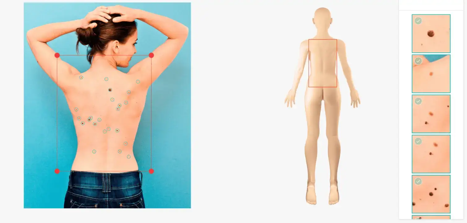
Mole mapping enhances the clinical examination by adding a detailed photographic record of your skin. The system:
- captures high-quality images of your skin
- records the location of individual moles
- creates a digital map for future comparison
This allows your skin to be monitored objectively over time.
How this differs from standard photography
Traditional mole mapping relies on basic photographs and manual comparison. AI-assisted mole mapping provides a more advanced approach by:
- systematically documenting all visible moles
- mapping their exact positions on the body
- allowing precise comparison between visits
The system is designed to ensure that even patients with many moles can be monitored consistently and accurately.
| Standard photography | AI-assisted mole mapping |
|---|---|
| Basic photographs | Systematically documents all visible moles |
| Manual comparison | Maps their exact positions on the body |
| — | Allows precise comparison between visits |
Layered imaging for greater detail
In addition to full-body imaging:
- The system captures images of all visible moles for baseline documentation
- The dermatologist identifies any moles that require closer assessment
- These selected moles are then photographed using high-resolution dermoscopic imaging, allowing detailed visualisation of structures beneath the skin surface
This combination provides:
- a comprehensive overview of the entire skin
- detailed analysis of clinically important lesions
Detecting change over time
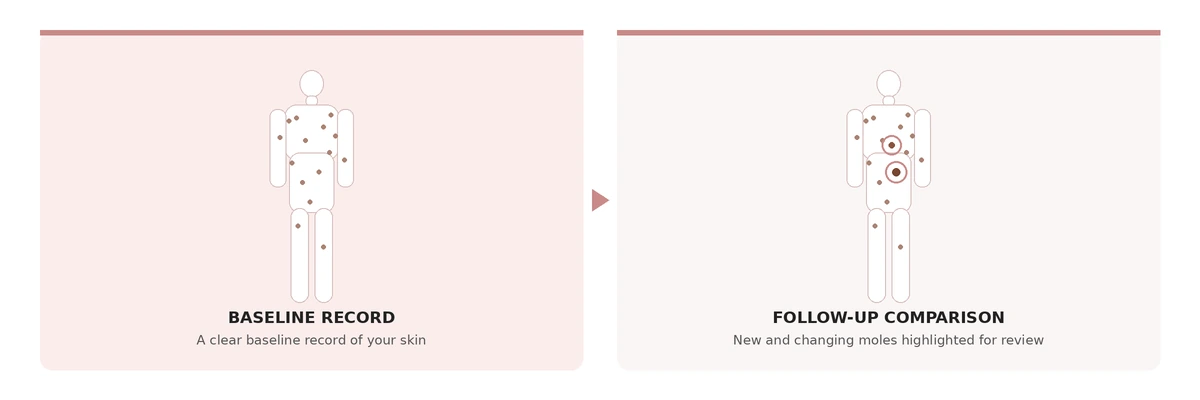
One of the key advantages of AI-assisted mole mapping is the ability to compare images over time. The system helps to:
- identify new moles
- detect subtle changes in existing moles
- highlight areas that may require closer review
This supports earlier detection of concerning changes that may not be obvious on routine examination alone.
Why this matters
Earlier detection of skin cancer
Skin cancers, including basal cell carcinoma, squamous cell carcinoma and melanoma, often begin with subtle changes. Accurate tracking improves the likelihood of detecting these changes at an early stage.
More precise monitoring
By documenting and following moles over time:
- stable lesions can be safely observed
- unnecessary procedures can often be avoided
Greater reassurance
Patients benefit from:
- a clear baseline record of their skin
- objective comparison at each visit
- a structured and consistent monitoring approach
Who should consider this?
AI-assisted mole mapping is particularly useful for individuals who:
Have multiple moles
Have atypical moles
Have a personal or family history of skin cancer
Are concerned about changing lesions
Want a more detailed and comprehensive skin check
Summary
A high-quality mole check combines expert clinical examination with advanced imaging technology.
At our clinic:
- every patient receives a thorough full-body examination by a dermatologist
- all visible moles are documented using advanced imaging
- selected moles are examined in greater detail using dermoscopic photography
- changes are tracked accurately over time
This integrated approach provides a more precise, consistent, and reliable way to monitor your skin compared to standard photography alone.
Frequently Asked Questions
What happens during my appointment?
All mole checks begin with a full skin examination by a dermatologist. You will be asked to undress to your underwear so the skin can be examined from head to toe, including less visible areas. A dermatoscope is used throughout to assess the structure of moles in detail.
Does mole mapping replace the doctor's examination?
No. Mole mapping enhances the clinical examination by adding a detailed photographic record of your skin. The clinical assessment remains the most important part of any skin check.
How is my skin monitored between visits?
The system captures high-quality images, records the location of individual moles, and creates a digital map for future comparison. This allows new moles to be identified, subtle changes in existing moles to be detected, and areas needing closer review to be highlighted.
Are all my moles photographed in detail?
The system captures images of all visible moles for baseline documentation. The dermatologist then identifies any moles requiring closer assessment, and those selected moles are photographed using high-resolution dermoscopic imaging.
Who benefits most from AI-assisted mole mapping?
It is particularly useful for individuals with multiple or atypical moles, those with a personal or family history of skin cancer, anyone concerned about changing lesions, and those wanting a more detailed and comprehensive skin check.
Book a mole check
If you are considering a mole check or skin screening, it is important to choose a clinic where both clinical expertise and technology work together.
Dr Ho has extensive experience in skin cancer detection, having practised dermatology in London where she performed skin cancer surveillance and surgery in Caucasian patients, and later as a Consultant Dermatological Surgeon at the National Skin Centre in Singapore treating skin cancers in Asian patients. This experience is important because skin cancers can present differently across skin types and may be easily mistaken for harmless moles or age spots.
Combined with advanced AI-assisted mole mapping, this allows for more accurate detection, monitoring, and early diagnosis.
Regular full-body skin checks, together with good sun protection and self-monitoring, remain the most effective way to detect skin cancer early, when treatment is simplest and outcomes are best.